AtaGenix Laboratories

Home - Support Center - Publication Insights

Small-Molecule DEPTAC TP2 Selectively Reverses Tau Hyperphosphorylation and Restores Cognition in Tauopathy Mouse Models

Release time: 2026-09-10   View volume: 5

Research Spotlight · Tau Pathology

Alzheimer's disease (AD) and a broader class of neurodegenerative disorders known as tauopathies are pathologically defined by the abnormal aggregation of hyperphosphorylated Tau protein (p-Tau) into neurofibrillary tangles (NFTs) within neurons. Under normal conditions, Tau stabilizes microtubules and supports axonal transport. Once its phosphorylation balance is disrupted, Tau detaches from microtubules, misfolds, and aggregates — triggering synaptic dysfunction and neuronal death. Over the past several decades, numerous Tau-targeted therapeutic strategies have been proposed, yet few have translated into clinical benefit. A shared limitation across these approaches is the lack of a treatment paradigm that can selectively clear pathological phosphorylated Tau while preserving its normal physiological function. Building on earlier work, a research team at the School of Basic Medicine, Huazhong University of Science and Technology (HUST) previously introduced the concept of DEPTAC (DEPhosphorylation TArgeting Chimera). Unlike PROTACs, which are designed to eliminate total protein levels, DEPTAC recruits endogenous phosphatases to the vicinity of Tau, directing them to selectively remove pathological phosphorylation marks without altering the phosphatase's overall activity or the function of healthy Tau. The team's earlier peptide-based DEPTAC molecules had already validated this approach across multiple preclinical models.

Building on that foundation, this study reports the redesign of the peptide-based molecule into a fully synthetic small molecule — TP2, a small-molecule DEPTAC that recruits the PP2A-Bα subunit.

Research Institution: School of Basic Medicine, Huazhong University of Science and Technology
Molecular Design and Synthesis

The researchers identified a pyrrolo[2,3-c]pyridine derivative (compound 11-5, derived from US patent US10022461) from the BindingDB database as the Tau-binding moiety. For the PP2A-recruiting moiety, they selected a phenothiazine-based scaffold (structurally related to perphenazine) previously validated to activate PP2A. The two moieties were linked via a PEG linker matching the length used in the earlier peptide-based DEPTAC. Through a sequence of reactions — substitution, nitro reduction, Ullmann coupling, and amide condensation — the team synthesized the final bifunctional molecule, TP2, with purity confirmed by HPLC to exceed 98%.
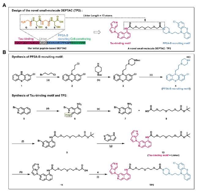
Design and synthesis of TP2
Figure 1. Design and synthesis of TP2
TP2 Simultaneously Binds Tau and PP2A, Inducing Formation of a Ternary Complex

The researchers confirmed this mechanistic hypothesis using three independent methods:

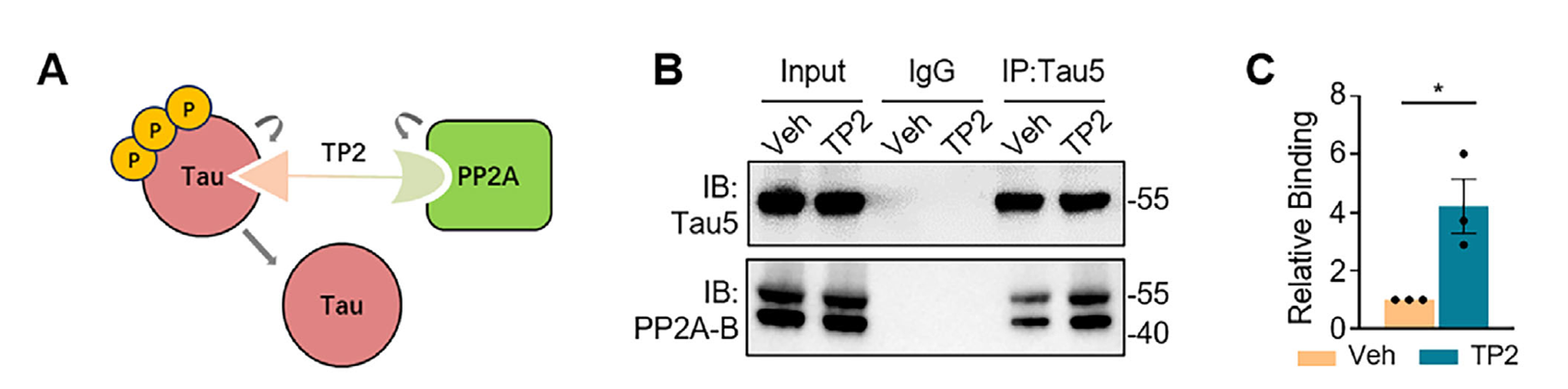
  • Co-IPCo-immunoprecipitation: In primary neurons, immunoprecipitating endogenous Tau with a Tau5 antibody after TP2 treatment revealed a marked increase in co-precipitated PP2A regulatory subunit.
    Assessing the interaction between endogenous Tau and PP2A-Bα by Co-IP
    Figure 2. Assessing the interaction between endogenous Tau and PP2A-Bα by Co-IP
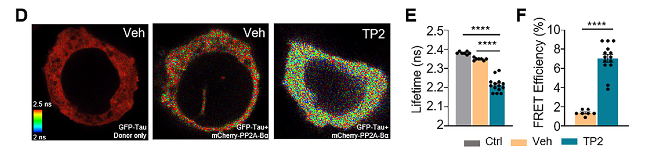
  • FLIM-FRETLive-cell imaging: In cells co-expressing GFP-Tau and mCherry-PP2A-Bα, TP2 treatment significantly shortened the donor fluorescence lifetime and increased FRET efficiency, indicating that the two proteins were brought within 10 nanometers of each other.
    FLIM-FRET experiments performed in live HEK293 cells
    Figure 3. FLIM-FRET experiments performed in live HEK293 cells to detect TP2-induced proximity between Tau and PP2A-Bα
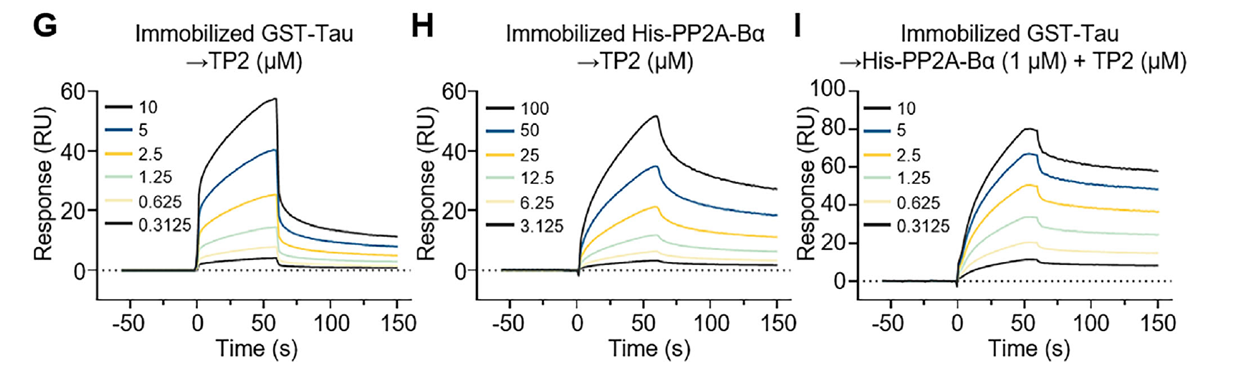
  • SPRSurface plasmon resonance: The dissociation constant (KD) of TP2 for GST-Tau was 7.36 μM, compared with 92.5 μM for His-PP2A-Bα — indicating substantially higher affinity for Tau. When 1 μM PP2A-Bα was added to the system, TP2's apparent affinity for Tau increased roughly threefold (KD dropped to 2.44 μM), demonstrating that the three components can form a cooperatively stabilized ternary complex.
    SPR characterization of TP2's direct binding to its targets and TP2-induced ternary complex formation SPR data 2
    Figure 4. Surface plasmon resonance (SPR) characterization of TP2's direct binding to its targets and TP2-induced ternary complex formation

In vitro microtubule polymerization assays showed that TP2 alone neither promotes microtubule assembly nor alters the kinetics of Tau-induced assembly, indicating that TP2 binding does not interfere with Tau's normal physiological function.

TP2 Promotes Tau Dephosphorylation and Reduces Neurotoxic Injury in Primary Neurons

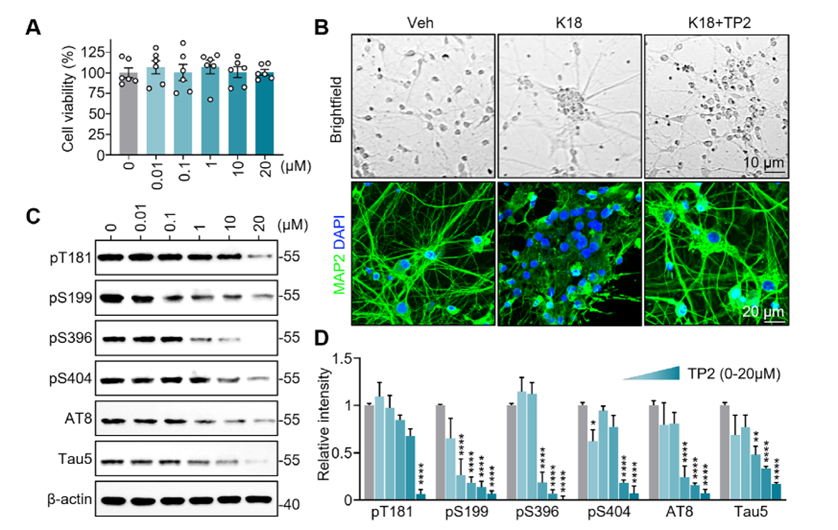
In primary rat hippocampal neurons, TP2 reduced levels of multiple phosphorylation sites (pT181, pS199, pS396, pS404, AT8) in a dose-dependent manner, with total Tau (Tau5) also showing a modest decline. CCK-8 viability assays confirmed that TP2 did not affect neuronal survival within the effective concentration range. The researchers then used K18 fibrils (an aggregation-prone fragment of the Tau microtubule-binding repeat domain) to induce Tau pathological toxicity: neurons exposed to K18 fibrils developed pronounced somatic aggregation and neurite dystrophy, while TP2 treatment ameliorated this phenotype and preserved a more intact dendritic network.

TP2 effectively reduces Tau phosphorylation and rescues K18-induced neurotoxicity in primary neurons
Figure 5. TP2 effectively reduces Tau phosphorylation and rescues K18-induced neurotoxicity in primary neurons

Compared with the previously reported peptide-based DEPTAC D16, TP2 achieved comparable dephosphorylation at a lower effective concentration and sustained its effect for longer after drug washout, demonstrating greater intracellular potency and a more durable duration of action.

Systemic Administration Delivers TP2 to Brain Tissue and Improves Pathology in Two Tauopathy Mouse Models

Pharmacokinetic data showed that TP2 was still detectable in thoroughly perfused brain tissue 30 minutes after a single intravenous injection (2.5 mg/kg), confirming that systemically administered TP2 reaches the brain. Building on this, the researchers administered seven intravenous doses to two model systems:

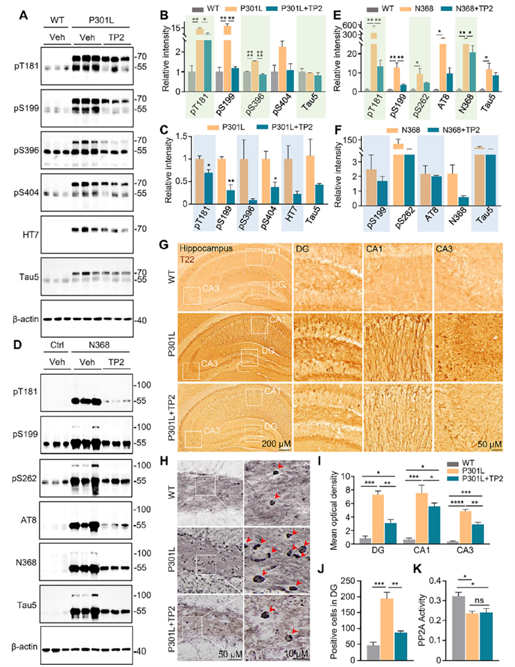
P301L mice (a mature-tangle model) and AAV-hTau-N368 mice (a fragment-toxicity model): TP2 significantly reduced pathological phosphorylated Tau at multiple sites in both models, while total Tau levels showed no substantial decline. This reflects the core feature of the DEPTAC strategy — correcting the phosphorylation imbalance rather than indiscriminately clearing Tau. TP2 was equally effective against already-aggregated insoluble Tau, oligomeric Tau, and neurofibrillary tangles, indicating that its effect is not limited to early-stage disease.

Key safety evidence: Overall PP2A phosphatase activity in the brains of P301L mice was unchanged before and after TP2 treatment. This shows that TP2 works by directing the phosphatase specifically to Tau, without altering the phosphatase's overall activity throughout the body — a key safety distinction from earlier phosphatase-activating therapies, which were often limited by systemic toxicity.
TP2 effectively reduces hyperphosphorylated, oligomeric, and aggregated Tau
Figure 6. TP2 effectively reduces hyperphosphorylated, oligomeric, and aggregated Tau, improves outcomes in tauopathy models, and leaves global PP2A activity unchanged
Concurrent Improvements in Neuronal Structure and Synaptic Function

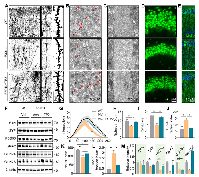
Beyond clearing pathological Tau, TP2 also repaired the neuron's structural "hardware." In P301L mice, TP2 reversed neuronal loss in the hippocampal CA3 region and restored dendritic branching complexity and spine density in CA1 pyramidal neurons, with MAP2 staining confirming the integrity of dendritic structure. Under finer-resolution electron microscopy, TP2 also reversed the decline in synaptic density, and key pre- and post-synaptic proteins (SYP, PSD95, GluA2, GluN2B) showed a trend toward recovery. Microtubule fragmentation was likewise improved, indicating that once Tau is dephosphorylated, its native microtubule-stabilizing function is restored in parallel.

TP2 treatment rescues neuronal morphology, synaptic integrity, and cytoskeletal structure in P301L mice
Figure 7. TP2 treatment rescues neuronal morphology, synaptic integrity, and cytoskeletal structure in P301L mice
Improvements in the Neuroimmune Microenvironment

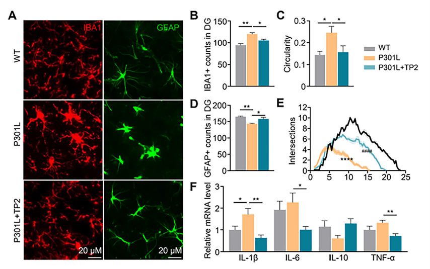
Tau pathology is commonly accompanied by neuroinflammation. Microglia in the dentate gyrus of P301L mice showed clear signs of activation (increased numbers, altered morphology); TP2 treatment significantly reduced microglial numbers and restored a resting-state morphology. Astrocytes told a different story: rather than the proliferative response typically seen, these 12-month-old mice showed astrocyte atrophy — reduced numbers and simplified processes — consistent with observations in human AD brain tissue. TP2 treatment reversed this atrophy, restoring both cell density and morphological complexity. qPCR results supported these findings: TP2 significantly reduced expression of the pro-inflammatory cytokines IL-1β, IL-6, and TNF-α, while the anti-inflammatory cytokine IL-10 trended upward without reaching statistical significance.

Effect of TP2 treatment on microglial activation and astrocyte atrophy in P301L mice
Figure 8. Effect of TP2 treatment on microglial activation and astrocyte atrophy in P301L mice
Recovery of Cognitive Function

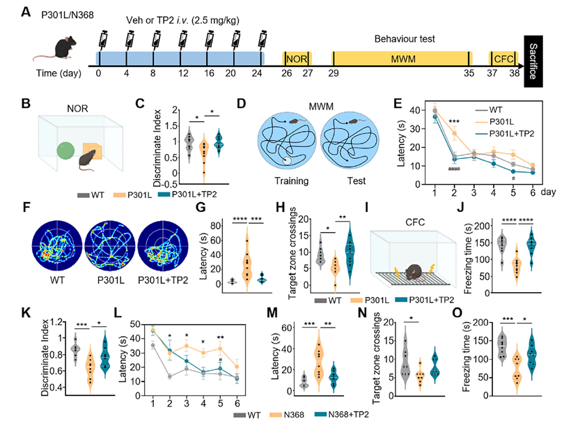
Having validated TP2's effects at the molecular, cellular, and structural levels, the researchers assessed functional outcomes using three behavioral tests:

  • NORNovel object recognition: TP2-treated mice recovered discrimination indices close to wild-type levels;
  • MWMMorris water maze: TP2 significantly shortened escape latency and increased target-quadrant platform crossings in both P301L and N368 mice;
  • CFCContextual fear conditioning: TP2 significantly increased freezing time, indicating restored consolidation of fear memory.

Consistent cognitive improvement across two mechanistically distinct models — the mature-tangle P301L model and the fragment-induced AAV-N368 model — provides strong support for TP2's therapeutic potential.

TP2 administration improves cognitive deficits in P301L and AAV-hTau-N368 mouse models
Figure 9. TP2 administration improves cognitive deficits in P301L and AAV-hTau-N368 mouse models
TECHNICAL SUPPORT · ATAGENIX CUSTOM ANTIBODY SERVICES

AtaGenix custom-produced the anti-Tau-N368 antibody used in this study for Western blot, enabling specific detection of the truncated N368 Tau fragment expressed in the AAV-hTau-N368 model — a key detection tool for characterizing this model's pathology and evaluating changes in the N368 fragment and its phosphorylation status following TP2 treatment.

The AAV-hTau-N368 model was one of two key animal models used in this study to validate TP2's broad-spectrum efficacy, recapitulating the toxicity induced by Tau fragmentation seen in sporadic AD. Because this model expresses an artificially truncated Tau fragment rather than full-length Tau, conventional antibodies struggle to detect it accurately and specifically — which is precisely where AtaGenix's custom antibody expertise adds value.

Thank You for Your Interest and Support!

AtaGenix remains committed to providing high-quality, end-to-end custom antibody production, protein expression, and related technical services,
helping drive the next generation of scientific breakthroughs.

Send a Message / Request a Service Plan
Messages
captcha